


$423.13
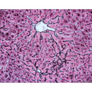
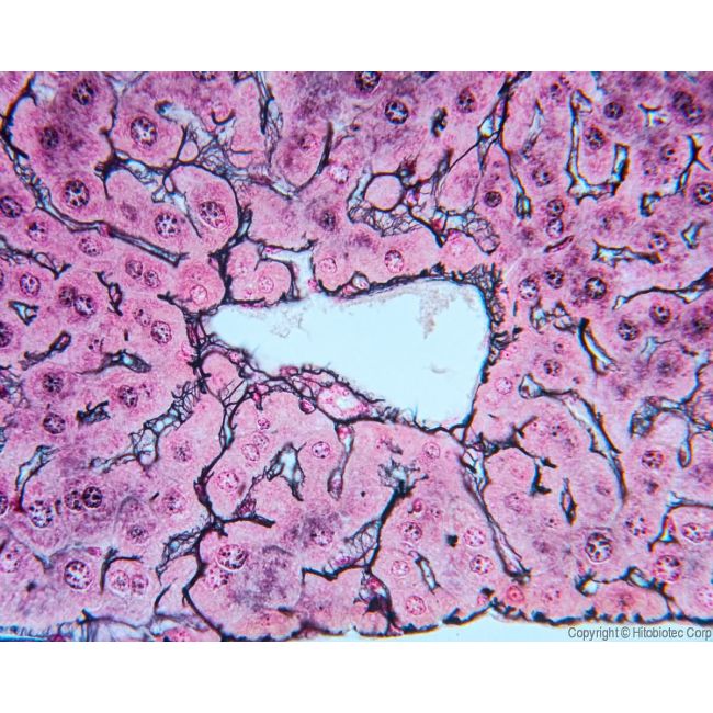
The reticular fiber silver stain is widely used to demonstrate reticular fibers. While the Gordon and Sweet method improved reticulin staining, traditional protocols often require large volumes of reagents, are costly and time‑consuming, and can be difficult to reproduce consistently.
The Hito Reticulin OptimStain™ Kit solves these challenges with simplified, user‑friendly procedures. Slides can be processed in small volumes, and the staining step can be controlled slide by slide for optimal staining and differentiation. When used properly, the kit delivers stable, improved quality with minimal overstaining, background, and artifacts.
Extensively tested on liver, spleen, pancreas, lung, and other tissues across multiple species, this kit provides sensitive visualization of the morphological details of reticular fibers. It is a simple, reliable solution for histological research.
SKU
HTKCS0102P
Write Your Own Review
Check items to add to the cart or